

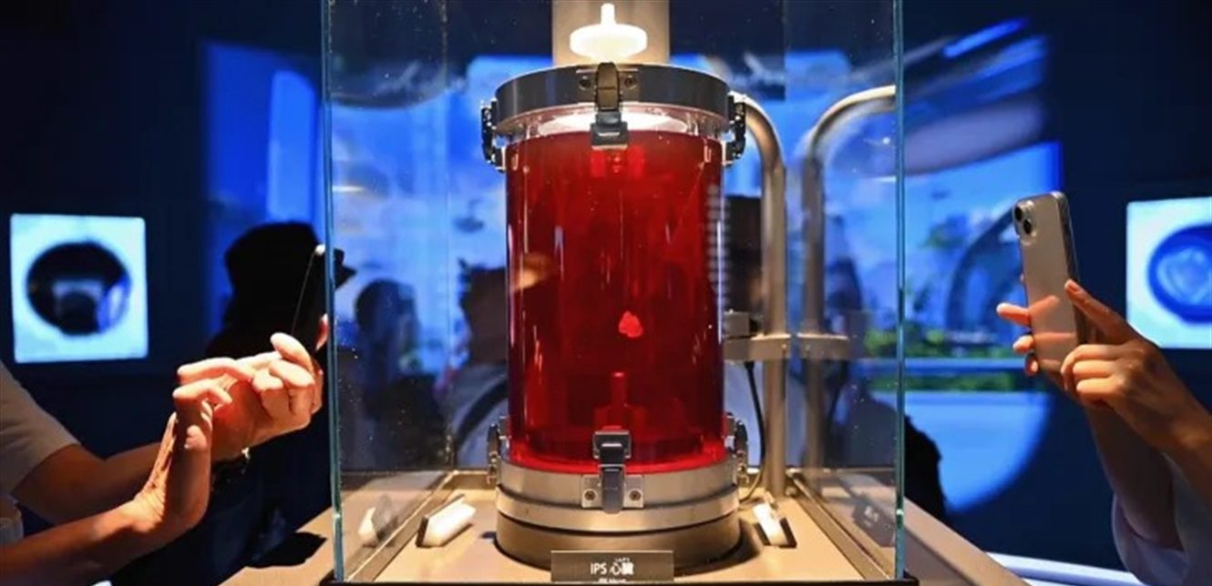
In a historic achievement, a research team from Osaka University in Japan announced its success in developing a miniature heart the size of a ping pong ball that beats independently, using stem cells taken from the patient’s own tissues.
Credit for this achievement goes back to 2012, when Japanese scientist Shinya Yamanaka won the Nobel Prize for his discovery of the technique of converting ordinary cells into induced stem cells, capable of transforming into any type of body tissue.
This discovery paved the way for an ambitious project later led by Professor Sawa Yoshiki of Osaka University, who devoted years of his research to achieving the dream of a “self-transplanted heart.”
The process begins by taking a small sample of heart cells, which are then reprogrammed to become induced stem cells. These cells are then directed via precise biological signals to become cardiac muscle cells, and then assembled into a three-dimensional structure that mimics real heart tissue. This tissue is placed in a warm solution rich in nutrients, and begins to beat on its own, as if it were a small heart forming inside the laboratory.
Thousands of patients die each year waiting for a suitable donor or suffering from the body’s rejection of the transplanted heart. But thanks to this technology, the heart can be made from the patient’s own tissue, eliminating the risk of immune rejection and making the transplantation process safer and more sustainable.
Currently, CureApp, managed by Sawa Yoshiki, is conducting clinical trials to transplant patches of self-heart tissue into the chests of patients. Initial results have shown that these patches act as “living bandages” that activate the weakened heart muscle and help it restore some of its vital functions.
Despite the great progress, the road is still long between a heart the size of a small ball and a complete transplantable heart. The most prominent risks lie in the possibility of the transplanted cells turning into cancerous cells if they are not carefully monitored, in addition to the difficulty of achieving compatibility in the electrical rhythm between the transplanted and natural heart, which could lead to serious disorders.
Scientists are also working on developing networks of fine blood vessels within the heart tissue using 3D bioprinting, in order to ensure the flow of blood and oxygen naturally within the new organ.
Although mechanical hearts and ventricular assist devices have saved the lives of thousands, they remain merely rigid machines that require maintenance and replacement. The heart cultured from the patient’s cells, on the other hand, is a living organ that grows and adapts to the body, giving hope for the birth of a new heart from the body itself, and not from another body.
With this discovery, a new page is opened in the field of regenerative medicine, where replacing damaged organs with cell transplants becomes possible. Today a heart the size of a ping pong ball… and tomorrow, perhaps a complete human heart born in the laboratory — vibrant, from man and for him.